St. Luke's Health joins CommonSpirit.org soon! Enjoy a seamless, patient-centered digital experience. Learn more

Imaging Department at Memorial Hospital - Livingston, TX
Mon
8:00 AM - 5:00 PM
Tue
8:00 AM - 5:00 PM
Wed
8:00 AM - 5:00 PM
Thu
8:00 AM - 5:00 PM
Fri
8:00 AM - 5:00 PM
Sat Closed
Sun Closed
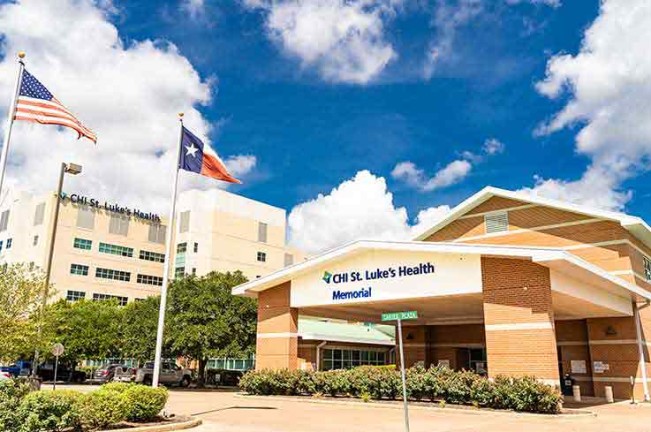
Located within St. Luke's Health-Memorial Livingston, the imaging department is a comprehensive, state-of-the-art imaging center servicing those patients staying in patient rooms, coming into the Emergency Department or needing outpatient imaging exams.
St. Luke's Health-Memorial Livingston allow patients to stay close to home for most of their diagnostic studies, including Magnetic Resonance Imaging (MRI), 128-Slice Computed Tomography (CT), 4D Ultrasound, 3D Mammography, Bone Density, Cardiac Imaging, Nuclear Medicine and Diagnostic Radiography (X-ray and Fluoroscopy).
The Livingston Imaging Department is an all digital, film-less imaging center, utilizing the Picture Archiving and Communication System (PACS) to deliver images for interpretation and storage. Allowing for a faster report turnaround time, PACS offers physicians the opportunity to consult patients on their results much sooner.
Scheduling and admission is easy. For outpatient studies, you or your physician can schedule your appointment. Either fax your physician orders prior to exam day or bring them with you when you register. The admissions desk is located in the front lobby of the hospital. Give the registration clerk your name and take a seat until the next clerk is available. After signing all applicable forms, a member of our guest services team will escort you to the imaging department.
Patients who are temporarily staying at St. Luke's Health-Memorial Livingston may be brought to the department from their room. However, some equipment, such as Ultrasound and X-ray, is mobile and exams can be performed in the patient rooms if needed.
MRI, CT, Ultrasound and Diagnostic Radiography technicians are always available in the event of an emergency.
Below is a brief description of each modality.
Utilizing advanced breast tomosynthesis – or 3D – technology, Hologic Genius™ 3D exams are clinically proven to significantly increase the detection of breast cancers, while simultaneously decreasing the number of women asked to return for additional testing.
In conventional 2D mammography, overlapping tissue is a leading reason why small breast cancers may be missed and normal tissue may appear abnormal, leading to unnecessary callbacks. A 3D mammography exam includes a three-dimensional method of imaging that can greatly reduce the tissue overlap effect.
During the 3D portion of the exam, an X-ray arm sweeps in a slight arc over the breast, taking multiple images. A computer then converts the images into a stack of thin layers, allowing the radiologist to review the breast tissue one layer at a time. A 3D mammography exam requires no additional compression and takes just a few seconds longer than a conventional 2D breast cancer screening exam.
Large clinical studies in the U.S. and Europe have demonstrated the positive benefits of a Genius 3D Mammography™ exam. The largest study to date on breast cancer screening using the Genius exam was published in the Journal of the American Medical Association (JAMA). Findings include:
Magnetic Resonance Imaging (MRI) uses a strong magnetic field and radio waves to align the hydrogen atoms in the body to see internal organ and tissue images without the use of radiation. These images show the difference between normal and diseased tissue enabling physicians to diagnose abnormalities.
MRI is a valuable tool to diagnosis conditions in the body including cancer, heart and vascular disease, stroke, breast disease, and joint and musculoskeletal disorders. Physicians use MRI scans to define anatomy and evaluate abnormalities related to head trauma, brain aneurysms and tumors, spinal cord trauma, glands and organs within the abdomen, and the structure of joints, soft tissues and bones.
Patients with a pacemaker, aneurysm clips or metal implants cannot be scanned due to the strong magnetic pull associated with the MRI. Some patients may experience claustrophobic feelings when their head is positioned inside the MRI bore. If you experience claustrophobia, please consult your physician prior to your exam date about taking a mild sedative.
The 128-Slice Computed Tomography (CT) uses x-rays to obtain multiple images from different angles around the body. As the fastest CT machine in the area, the noninvasive, painless medical test helps physicians diagnose and treat various medical conditions such as kidney, lung, liver, spine, and blood diseases, cancer, tumors and cysts, as well as blood clots, hemorrhages and infections.
While CT uses X-ray technology, it is distinguished from other diagnostic imaging tools like traditional X-ray and MRI by its ability to display a combination of soft tissue (like muscles, tissue, organs and fat), bones and blood vessels all in a single image. CT imaging is useful because the tissue is seen with great clarity, making disease diagnosis much easier.
A computer processes this information to create an image showing body tissues and organs in cross-sectional views called slices. Imagine the body as a loaf of bread and you are looking at one end. As you remove each slice of bread, you can see the details of the next slice. CT scan slices are similar allowing physicians to see small areas from different views.
The Livingston Imaging Department's 128-slice CT scan produces results in just four minutes and gives physicians a clearer image than ever before.
Patients who are unable to complete a MRI study, due to pacemaker, defibrillator or claustrophobia, can have a CT exam. Depending on the type of CT scan ordered, you may be asked to modify your diet. Some exams require that you drink a contrast material prior to your exam. Other exams require a contrast material injection, usually in a vein in the arm. The study usually takes about 20 minutes to perform.
Ultrasound, or sonography, uses high frequency sound waves to produce images of the fetus and internal organs.
The state-of-the-art 4D imaging system is used to help monitor and diagnose conditions in many parts of the body. Sound waves are transmitted by a transducer through the patient and reflected back when reaching the area being examined. Areas that are monitored are the growth of an unborn child, as well as arteries, veins, liver, gallbladder, pancreas, thyroid gland, lymph nodes, ovaries, testes, kidneys, bladder and breast. Abnormal lumps in one of these organs can be determined as a tumor or fluid-filled cyst. The imaging system also assists in diagnosing gynecological issues by allowing physicians the capability to study the uterine cavity, the fallopian tubes and the ovaries in detail. It also assists in the detection of ectopic pregnancies, ovarian cysts and endometrial polyps. The 3D and 4D capabilities allow for enhanced breast imaging and elastography, a non-invasive method of detecting tumors.
Because it can be used in the most delicate conditions without major side effects, ultrasound has become one of the most popular diagnostic methods among both patients and physicians. It provides a safe, fast and relatively painless means of diagnostic imaging on an outpatient basis.
A bone mineral density test, also known as a DEXA (dual-energy x-ray absorptiometry) scan, is a non-invasive and painless method to determine your bone health. The test measures how many grams of calcium and other bone mineral content are within a certain segment of bone. The higher your mineral content, the denser and stronger your bones are and less likely to break. Doctors use a bone density test to identify osteoporosis, determine your risk for fractures and monitor response to osteoporosis treatment.
Bone density is measured in the spine, wrist, and hip. These locations are the most likely to break because of osteoporosis.
The results from the scan will help your physician determine if medical supplements are necessary to increase your bone density.
Post-menopausal women over the age of 65 are recommended to have a bone density test.
The radiation exposure is minor (less than someone receives with a chest x-ray).
A Nuclear Medicine exam is a diagnostic procedure that uses a radioactive tracer substance and a special camera to image body and organ anatomy and function non-invasively. Livingston was one of the first hospitals in the state to obtain the Symbia® S Nuclear Medicine system with high definition digital detectors that offer exceptional imaging performance and expanded clinical capabilities. Tumors, infection and other disorders can be detected by evaluating organ function. Nuclear Medicine is used to:
Nuclear Medicine has become a very popular method of diagnosing and following coronary artery disease without the risk involved with cardiac catheterization. Nuclear medicine is the primary way metastatic bone disease is diagnosed.
Some scans require certain preparations prior to the appointment. Procedures involving evaluation of the stomach require the patient to arrive at the appointment with an empty stomach (NPO). Another procedure evaluating the kidneys requires the patient to drink plenty of water before the test. Your physician or the imaging center staff can give you preparation instructions for your exam.
Diagnostic radiography, also known simply as X-ray, is the oldest, most frequently used form of medical imaging. It is widely used to identify healthy and abnormal conditions in the body. X-ray imaging is fast, easy and painless. It is useful in the diagnosis and treatment of bony and soft tissue injuries, infections, and fractures. Common x-ray exams are:
Fluoroscopy uses a continuous X-ray beam to create a sequence of images that are digitally transmitted to a high-resolution TV monitor. The body part and its motion can be seen in “real time” detail.
Fluoroscopy enables physicians to look at many body systems, including the skeletal, digestive, urinary, respiratory, and reproductive systems. Fluoroscopy is used to evaluate specific areas of the body, including the bones, muscles and joints, as well as solid organs such as the heart, lung or kidneys. Examinations and procedures that use fluoroscopy along with preparations are:
Diagnostic radiography does involve some exposure to radiation. However, special care is taken during the exam to minimize exposure and maximize safety for the patient by using lead aprons or shields to block radiation when needed during the exam. The radiation dose for diagnostic radiography is about the same as the average person receives from everyday background radiation in about 10 days.
Always consult your physician on the detailed instructions to prepare for a test. Inform your physician of any medications you are taking and any allergies, especially to contrast materials. Some medications may be taken prior to an exam and others may negatively interact with your results.
Looking for a doctor? Perform a quick search by name or browse by specialty.