St. Luke's Health joins CommonSpirit.org soon! Enjoy a seamless, patient-centered digital experience. Learn more

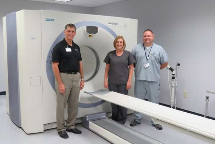
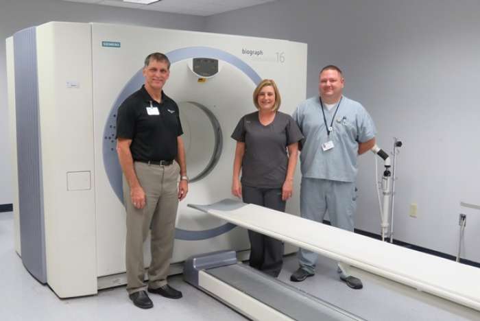
In the fight against cancer, patients deserve the best when it comes to a diagnosis and treatment, and it takes a team of caring, trained professionals to achieve the best results. As one of approximately 800 PET/CT certified technologists in the nation, Bill Schelling, CNMT, PET, and R.T. (CT), brings a wealth of experience to the East Texas area identifying cancerous tumors at St. Luke’s Health Memorial Lufkin.
A PET/CT scan combines the imaging capabilities of a Positron Emission Tomography (PET) and a Computed Tomography (CT) to help physicians locate cancerous cells in the body to determine treatment. It can also be used for brain imaging to diagnose Alzheimer’s and epilepsy.
Schelling received an Associates of Applied Sciences in Nuclear Medicine from Southeast Technical Institute in Sioux Falls, SD in 2006, which included PET training. He also attended CT training at Galveston College last summer. He is board certified in PET, nuclear medicine and CT.

“All I did was PET/CT scans where I worked before, and we were very busy,” said Schelling, who has worked as an imaging technologist for nine years. “I’ve done approximately 8,000 PET/CT scans in my career. Receiving my PET scan accreditation means I’ve put in the extra work. It shows that I’ve done my training and have conducted numerous scans.”
A PET scan is a highly sensitive nuclear medicine exam that detects signs of actively growing cancer cells. A CT scan provides the internal anatomical detail of the location, size and shape of abnormal cancerous growths. Alone, each imaging test has particular benefits and limitations. But when the two images are fused together, PET/CT provides complete information on cancer location and metabolism.
“The PET/CT shows just how active the tumor is, and the oncologist can then tailor the patient’s treatment,” Schelling said. “This is a much more active scan than just a picture; it’s more like we’re watching a movie than looking at a still picture. Because it is so accurate there is no better scan for cancer imaging than the PET/CT.”
Patients are injected with a radioactive glucose tracer about 45 minutes prior to the scan. The dye accumulates in areas of the body that are metabolically active such as some organs and cancerous tumors. The PET and CT scans are then simultaneously performed. Together the scans create a color image that allows physicians to see “hot spots” where cancer lives.
It is unnecessary for cancer patients to travel outside of the deep East Texas area to receive a PET/CT scan when Memorial provides such high skilled technologists right here at home.
“Oftentimes, people don’t realize we have this technology right here in Lufkin,” said Amy Ford, R.T. (R), CNM, certified nuclear medicine technologist and certified X-ray technologist. “Our goal is to make people aware of what we have and who we have. Why should you go to Tyler or Houston when you can stay here for the same scan?”
Patients must be diagnosed with certain cancer indications prior to a physician ordering a PET/CT scan.
“CHI St. Luke’s Health Memorial invested in this technology to provide a clearer, more 3-dimensional image that greatly assists in cancer treatment planning,” said Director of Imaging Services and the Temple Cancer Center Bill Malnar, M.S., R.T. (R). “Patients are ultimately in charge of their cancer journey, and they can choose to start right here at Memorial knowing they are in good hands with such experienced technologists.”
The PET/CT equipment is located at the Temple Imaging Center on the St. Luke’s Health Memorial Lufkin campus. The equipment and the staff at the Temple Imaging Center are all accredited by the American College of Radiology.
For more information, call the Temple Imaging Center at 639-7374.
Publish date:
Wednesday, September 30, 2015Looking for a doctor? Perform a quick search by name or browse by specialty.